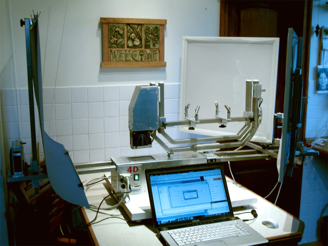
Interactive 4D image reconstruction scanner
Our company, with the help of 4D Anatomy LLC’s patent and the PIAC-13 grant fund supporting R&D projects, developed a special scanner in 2014 which helps doctors take rapid images of anatomical preparations systematically from all directions.
The 4D map that shows the details of the human body is no longer only a virtual simulation, but also a visual spatial reality with never before seen detail. In Hungary, due to the limited nature of autopsy training, this programme means an enormous help. The remarkably quick and at the same time very exact photography establishes the basis for a whole new teaching and demonstration method through which the realistic human body becomes available to anyone to study.
The entire human body is visible in never before seen detail: the Hungarian team developed a unique 4D scanner
Budapest, 26 January 2015 – The 4D Anatomy technology which has been further improved by the medical and engineering team of Four Point Kft. provides an entirely new view during autopsy and surgical simulations. The photos of anatomical preparations are taken from the most varied positions in space and at high speed with the help of a worldwide unique scanning robot. The Hungarian development not only aids the learning of university students, but also provides outstanding support for doctors in the detection of rare conditions.
Four Point Kft., with the help of 4D Anatomy LLC’s patent and PIAC-13 grant fund supporting R&D projects, developed a special scanner last year through which photos of anatomical preparations can now be taken systematically from all directions. Images are reconstructed with the 4D Anatomy software and converted into interactive 4D modules which allows moving and manipulation of the injured organ in 4D under the control of the user. The remarkably quick and at the same time very exact photography establishes the basis for a whole new teaching and demonstration method through which the realistic human body becomes available to anyone to study.
Although with the help of existing and practically used 3D CT and MR technologies, rotating scanned objects has already offered significantly more information compared to static imaging, unfortunately, the visual display has a drawn, artificial effect so they do not reflect colour, shade and material features at all. In contrast, the 4D map that shows the details of the human body is no longer a mere virtual simulation, but a visual spatial reality with never before seen detail.
“The uniqueness of the programme which was developed by Hungarians lies in the fact that, in addition to the anatomy of the human head and neck, it also displays other body parts with cutting edge technical solutions, unique precision and amazing detail. Small details of the human body can be displayed in 4D and studied both vertically and horizontally even from a home PC. The programme is also available from a cloud provider therefore it can truly be considered a 21st century teaching and presentation platform” – explained Dr László Kutor, robot designer, developer.
In Hungary, due to the limited nature of autopsy training, this programme means an enormous help. It improves the efficiency of medical diagnosis and is also very cost-effective because its application does not require the installation of new, high-value diagnostic tools in the already overburdened healthcare system.
“The quality of neurosurgery training is affected by a number of factors nowadays. Technical possibilities, economic development and cultural barriers may all limit access. The teaching of surgical manoeuvres, the design and performance of surgery require thorough preparation to keep errors to a minimum. Therefore there is an increased need for alternative methods and teaching tools which can help in the proper selection of surgical tools and procedure.” – emphasised Attila Balogh, MD, Neurosurgeon.